Physical therapy Techniques and Knowledge Sharing. https://mcqphysiotherapy.blogspot.com/
Friday, December 22, 2006
ROLE of Orthopaedic Physical therapist
• Orthopeadics & physiotherapy are Two faces of same coin.A Good Orthopedic Surgeon is one who has a Good Physiotherapist (John Ebenezer. 2003).
• Physiotherapist blends with orthopedics and makes patient put back to normal
( Pre injury) state.
• The role of Physiotherapist doesn’t starts after the fracture is fixed or after the disease is healed but it starts from the day one of the onset of Disease or Fracture.
Causes for weakness following Immobilization
• Trauma to soft tissue
• Surgery (e.g., joint replacement)
• Joint disease (e.g., osteoarthritis)
• Prolonged immobilization
• Neuromuscular disease
Ultimate Purpose of exercise program is
· To restore function
· To restore Performance
· To restore Muscle strength
· To restore endurance – Pre trauma level
ISOMETRICS
n Maintain Muscle Strength
n Done at the early stage
n Done with POP or Plaster cast
n Done in Immobilization period
ISOTONICS
· Increase Muscle Strength
· Done at the Intermediate stage
· PRE are advised
· Dumbbells, Weight cuff, sand bags are used.
ISOKINETICS
Joint movement at a constant rate
Improves Muscle Strength
Done at the Late stage
Cyberx is used to train.
CONDITIONING EXERCISE:
Increase endurance
Cardiopulmonary fitness increases
Conditioning exercise enhance peripheral oxygen utilization & efficiency & result in Aerobic muscle metabolism.
E.g Stationary bicycle, Treadmill
Chikungunya Facts
Also called chikungunya virus disease or Chikungunya fever,
Caused by Musquito Bite Named as Aedes aegypti is the major vector of chikungunya virus.
It is a viral illness that is spread by the bite of infected mosquitoes. The disease is characterized by severe, sometimes persistent, joint pain (arthritis), as well as fever and rash. It is rarely life-threatening.
Chikungunya occurs in Africa, India, and Southeast Asia.
1. There is no specific treatment for chikungunya.
2. Prevention centers on avoiding mosquito bites in areas where chikungunya virus may be present, and by eliminating mosquito breeding sites.
How soon after exposure do symptoms appear?
The time between the bite of a mosquito carrying chikungunya virus and the start of symptoms ranges from 1 to 12 days.
Diagnosis:
Chikungunya is diagnosed by blood tests. Since the clinical appearance of both chikungunya and dengue are similar, laboratory confirmation is important.
TREATMENT: There is no specific treatment for chikungunya., Only supportive therapy is given nonsteroidal anti-inflammatory drugs, and getting plenty of Rest.
Infected persons should be isolated from mosquitoes in as much as possible in order to avoid transmission of infection to other people.
PREVENTION
Use mosquito repellents on skin and clothing.
Use insect repellents that have been approved by the Environmental Protection Agency (EPA). They are safe and effective.
When indoors, stay in air-conditioned or well screened areas. Use bed nets if sleeping in areas that are not screened or air-conditioned. · When outdoors during times that mosquitoes are biting, wear long-sleeved shirts and long pants. Lasts through 5-6 washes.
Footwear modifications reduce load on knee.
Patients with Osteoarthritis Knee may benefit from footwear modifications that can effectively reduce knee joint moments. Foot wear design in which the Rear foot portion of the Mid sole moves independently of the rest of the shoe.
The design allows the foot to move mediolaterally as well as anteroposteriorly relative to the shoe outsole-which, the researchers found, can significantly reduce both abduction and external rotation moments at the knee.
Rigid midsoles were associated with significantly higher maximum knee abduction moments and external rotation moments than the softer midsoles.Footwear modifications that reduce knee joint moments may also be able to help patients with knee osteoarthritis
Clacaneus Valgus & Varus also contribute more stress on the knee will cause pain in Knee.
Alteration in Foot e.g. Altering foot by elevating foot wear medially at the sole cause marked reduction in stress implicated on the joint. Will make the patient feel better
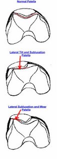
Chondromalacia Patella
The term chondromalacia patella has often been used interchangeably with patellofemoral syndrome and anterior knee pain,
The term chondromalacia patella describes the softening and progressive breakdown of the articular cartilage of the patella.
It occurs secondary to an underlying condition such as malalignment or trauma (Greenfield and Scott, 1994).
Grades of the extent of cartilage damage. Eisele (1991),
1. Grade 1 - Articular cartilage only shows softening or blistering
2. Grade 2 - Fissures appear in cartilage
3. Grade 3 - Fibrillation of cartilage occurs, causing 'crabmeat' appearance Grade 4 - Full cartilage defects are present and subchondral bone is exposed
DO BONE MINERAL DENSITY IMPROVES WITH EXERCISE
The intense exercise training program presented here was effective in improving strength, endurance, quality of life parameters, and even BMD in women in their critical early postmenopausal years, Kemmler W, et al., 2002.
Physical activity as a way to prevent osteoporosis is based on evidence that it can regulate bone maintenance and stimulate bone formation including the accumulation of mineral,
Strengthening muscles, Improving balance, and thus reducing the overall risk of falls and fractures.
These exercise-stimulated increases in bone diameter diminish the risk of fractures by mechanically counteracting the thinning of bones and increases in bone porosity.
Seven principles of bone adaptation to mechanical stress
Principles are
(i) Be dynamic, not static;
(ii) Exceed a threshold intensity;
(iii) Exceed a threshold strain frequency;
(iv) Be relatively brief but intermittent;
(v) Impose an unusual loading pattern on the bones;
(vi) Be supported by unlimited nutrient energy; and Include adequate calcium and cholecalciferol (vitamin D3) availability. Borer KT, et al., 2005
Wednesday, November 22, 2006
Foot Types.
Foot Types.
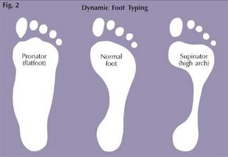
1. Fore foot varus – in this deformity the fore foot is inverted relative to the hind foot in subtalar neutral position (also known as metatarsal adductus or varus). Deviations of the fore foot greater than 5° will ultimately lead to foot pain and needs to be treated. The first metatarsal becomes hypermobile. To compensate and allow the fore foot to bear weight during walking, the subtalar and the midtarsal joints pronate. The hind foot is in valgus position.
2. Rear foot varus – in this deformity the posterior surface of the calcaneum is inverted in relation to the ground with the subtalar joint in neutral. As a result of this the plantar surface of the foot becomes inverted and the patient tends to walk on the lateral surface of the foot. During weight bearing, this is compensated by pronating the foot to bring the medial side to ground contact. The first ray is plantar flexed.
3. Fore foot valgus – this is where the fore foot is everted in relation to the calcaneum with the subtalar joint in neutral position. This causes the medial side of the foot to come in contact with ground very early in the gait cycle. The foot may compensate by dorsiflexing the first metatarsal joint (lower first metatarsal), to allow the lateral side to come in contact with the ground. If this movement is insufficient then the midtarsal and subtarsal joints may supinate
4. Rear foot valgus – this happens as compensation to fore foot varus. This may be defined as a deformity in which the heel is everted in relation to the midline of the leg with the subtalar joint in neutral. This causes pronation of the leg and internal rotation of the leg.
Osteoporosis
Osteoporosis
It is a generalised diseases of bone in which there is a marked decrease in the amount of Bone.
· Acc. To WHO Less than 2.5 SD consider as Osteoporosis.
· Common in Post menopausal women, due to decrease of Oestrogen
Common Sites are : neck of femur, proximal Humerus, proximal Tibia, Pelvis, metatarsal bones
Risk Factors:
· Age = Over 50 Yrs
· Sex = women > men
· Race = whites > blacks
· Body type = Small frame > large frame.
· Family history
· Post menopausal.
· Nutritional Factors
· Life style factors
· Medical factors
· Mensural cycle disturbance
Treatment:
· Exercise
· Oestrogen replacement
· Increase calcium consumption
· Weight bearing exercises
· Strengthening exercises
· Walking best exercise increases strength of Lower extremities and Spine
Geriatric Rehabilitation
Ø Geriatric Rehabilitation is the branch of rehabilitation dealing with alleviative or remedial, preventive or social aspects of the elderly disabled and disadvantaged.Geriatrics is the Branch of medicine dealing especially with the Problems of ageing and disease of the elderly
PRINCIPLES
Ø Ascertain level of function.
Ø Ascertain available resources and options
Ø Avoid im-mobilization
Ø Beware of altered physiological reactions
Ø Determine patients Goals & Motivation.
Ø Determine family expectation.
Ø Differentiate between delirium, dementia, depression.
Ø Emphasis function, management.
Ø Emphasis task specific exercises, simplify program
Ø Encourage socialization and stimulation
Ø Minimize medications
Ø Realize the function may not be regained, recognize that patients have multiple interacting impairments.Understand that improvement occurs in slow increment
OVERUSE INJURY
n Anatomical factors
n Excessive body weight
n Imbalance in muscle strength
n Improper shoe wear (specific to activity)
n Improper technique
n Pushing too hard (not allowing for rest)
n Repetition of exercises – no variety
n Training errors (too much, too soon)
n Training surfaces
n Training Climate
n Improper Warm up or Cool down Phase
Individual Joints involvement:
n Back: (low back pain syndrome) caused by anatomical, lack of stretching, imbalance of muscle strength, impact exercise
n Feet/Ankle: (Plantar Fasciitis, stress fractures, bunions, achilles tendonitis) caused by anatomical, improper shoes, lack of stretching, impact exercises.
n Hips: (Psoas bursitis) caused primarily from lack of stretching, improper technique, anatomical, overuse.
n Knees: (Chondromalacia, Patellar tendonitis) usually caused by running everyday, downhill running/walking and muscle imbalance.
n Legs: (Shin splints, Anterior compartment syndrome, Stress fracture) caused by lack of stretching, improper shoe wear, uneven surfaces, repetitive running, muscle imbalance between calves and shins (anterior tibialis)
n Shoulder/Arms: (Subacromial bursitis, Rotator cuff tendonitis) caused by overuse, lack of stretching, muscle imbalance, lifting too much weight, improper technique using weights.
Treatment
Treatment usually given is RICE.
n REST: Essential for healing
n ICE: Reduce pain and swelling
n COMPRESSION: Help to reduce swelling and provide support to injured area
n ELEVATION: Reduces pain and swelling
LATE RESPONSE
F- WAVE
l The F wave results from Antidromic activation of motor neurons involving conduction to and from spinal cord and occurs interface between the peripheral and central nervous system.
l Name attributed – Foot muscles. (Magladery & Mc Dougal 1950).
STIMULATION: Supramaximal from any distal muscles by stimulating appropriate nerve.
l 25 % above maximal.
l Not more than 0.5 Hz frequency.
l Electrode placed on a belly tendon montage similar to MNCV.
l Amplifier Gain 200- 500 μv.
l Sweep speed 5-10 ms / div.
l Slight voluntary contraction may enhance F waves.
l F- wave requires amplitude more than 20μv.
l Clinical purpose 10-20 F waves are adequate.
l For study persistent 20 responses desirable.
l Latency, chronodispersion, persistence and amplitude.
l F latency is related to Height, Limb length and Age.
l The correction for these improves sensitivity.
l F latency usually expressed as Minimal latency.
l F wave calculate NCV as well as Proximal conduction time.
l It refers to difference between minimal and maximal latency in a series of F wave.
l It measures range of conduction of F wave.
DURATION
l Tells the number of motor units used
AMPLITUDE
l Refers the Size of motor units
PERSISTENCE
l The Number of Occurrence of response divided by the number of stimuli.
l Refers to the Antidromic activity of a particular motor neuron POOL.
l F amplitude to the associate M is a measure of proportion of motor neuron pool activated by Antidromic stimulation.
l Mean is used to calculate FM ratio.
l Normal F latency in adult 31ms for Hand & 61 ms for Foot.
l Rt to Lt asymmetry 2ms in Hand & 4 ms in Foot.
l Chronodispersion of F wave for abductor pollicis brevis is 3.6+/- 1.2
l F wave amplitude is 5% of M wave.
l Persistence of F wave ab.dm is & adductor hallucis are 0.8 – 0.9
l F differ b/w neonates, child, and limb length.
l Elders have long latency than men.
l F latency in Men 31 ms and women is 34.4 ms
l The Latency can be sensitive measure in Polyneuropathy.
l Abnormal seen in distal motor conduction are unremarkable.
l GBS slowing of F wave.
l Abnormality seen in PNL, Radiculopathy, peripheral neuropathies.
l ALS reduced persistence
l Chondrodispersion increased in Polyneuropathies.Duration prolonged UMN lesions
ARE YOU FIT ?
ARE YOU FIT ?
Fitness is not something that belongs to the young – it’s everybody’s right, no matter what the age…
& How many of you feel that “fitness” is an important part of your job?
& How many of you work out more than 6 days a week?
& How many of you feel that management supports your “wellness”?
Why Fitness?
n Fitness improves overall health.
n An established fitness regime improves personal attitude.
n Helps decrease absenteeism and increase productivity in the workplace.
n Maintaining good physical fitness can improve work and cope up with stress.
In order to have an effective fitness program – you MUST balance all of the fitness components. …… No Short Cuts!
Program starts with
n Warm-Up ( 10—15 Mins )
n Aerobics (Exercise with O2)
n Dumbbell Exercise
n Flexibility (stretching)
n REST…….and Relaxation….. simply named Cool down ( 5—10 Mins)
Swiss ball exercises
The exercise ball (or Swiss ball or Physio ball) is a versatile piece of exercise equipment available to help people with pain. In particular, many Swiss ball exercises are designed to bring movement to the joint in a controlled manner to keep the joint nourished.
The exercise ball has additional applications in areas such as
General fitness,
Strength or weight training, and
Exercise for pregnant women.
Role of Swiss Ball on Low back pain :
Reducing Back Pain,
Strengthening Core Body Muscles
“The exercise ball - also called a Swiss ball or Physio ball - is a conservative treatment option for back pain sufferers and is designed to help prevent further episodes of low back pain as part of a rehabilitation program. The exercise ball is effective in rehabilitation of the back because it helps strengthen and develop the core body muscles that help to stabilize the spine.
The muscles used to keep in balance on the Swiss [exercise] ball become stronger. Individuals build strength of back muscles and abdominal muscles.
The benefits of Swiss ball are.
Improved muscle strength
Greater flexibility and range of motion of the spine
Enhanced balance and coordination of core muscle groups used to stabilize the spine and control proper posture while using the exercise ball
Increased tendency to maintain a neutral spine position during exercise
Exercise are simple and easy to do
Easy Home programme
Wanna to Know more Contact…………
Wednesday, November 08, 2006
PHANTOM PAIN
Phantom pain is described as "cramping, shooting, aching, hand clenched in a tight fist, toes out of joint, frozen or rigid joints, or any combination of these" and usually subsides within a year after surgery
WHY IT OCCURS ???
• Prior experience with pain prior to amputation
• Incorrect surgical procedure
• Climatic conditions
• Stress
• Inactivity
HOW TO TREAT IT ??????
• Psychological Counseling
• P.T Modalities like TENS, US, Massage
• Bandaging, Acupuncture.
• Regular exercise,
• Stretching.
Wanna know more ………………
Effect of ligaments and Muscles in Osteoarthritis
Effect of OA Changes in Ligaments
Abnormal joint alignment stresses
Effect of OA Changes in Muscles
Immobility shortens pain, causes guarding and reflex inhibition, leading to weakness
Effect of OA Changes in Bones
Subchondral bone remodeling changes shock-absorbing properties, joint-margin spurring leads to bony blockade and pain
Effect of OA Changes in Extra articular system
Increased energy expenditure from abnormal movement patterns
Effect of OA Changes in Synovium
Abnormal joint alignment stresses
Effect of OA Changes in Articular cartilage.
Thickening to softening, to thinning to loss
Wanna know more ………………
CAN YOU STRETCH YOUR NERVES ?????
The muscles, ligaments, tendons even bone has elastic properties and has elastic limit. But we still don’t know the exact limit of elasticity of Nerve. I have certain queries to find out the behavior of nerve when application of tensile stretch.
1. How much load does the nerve requires to cause a conduction block?
2. At what percentage of stretching cause rupture of nerve?
3. When the nerves will behave a plastic property?
4. After releasing of stretch does the nerve regenerate?
Wanna to learn more contact ………………..
TRANING EFFECT ON LIGAMENTS
Physical training has found to increase the tensile strength of ligaments,
Physical training produces considerable effect on ligaments.
Tipton et al., tries to find out the strength of MCL in dogs.
He concluded in his study that the strength of MCL develops in dogs as a result of training. The ligaments in the trained group are stronger and stiffer than the control group.
Immobilization found decrease strength in Ligaments--Aminel et al.,
Following immobilization when a load is applied to ligaments results in failure
Noyes et al., found there is about 39 % of decrease of stress tolerance in ligaments following immobilization
Immobilized ligaments displayed more elongation and significantly less stiff.
Following immobilization if ligaments are trained it need considerable time to develop its strength.
Aminel et al., found strength training to ligament injury has to be started as soon as possible to get back the ligament in normal
Wanna to learn more contact ………………..
Saturday, October 21, 2006
COMPUTER VISION SYNDROME
Symptoms:
ï Eyestrain
ï Blurred vision
ï Dizziness or nausea
ï Headaches
ï Red, dry or burning eyes
ï Increase in near sightedness
ï Change in color perception
ï Slow refocusing
ï Difficulty seeing clearly at a distance after prolonged computer use
ï Excessive fatigue
ï Eye-teaming problems and/or occasional double vision.
ï Slow refocusing when looking from screen to distance objects
Apart from these the professionals may experience Neck pain, Shoulder pain, Wrist pain, and Low back pain.
When you find any of these symptoms, then you are at a great risk of Having Computer vision syndrome.
Computer vision syndrome (CVS) is not a complicating disease, it is easily preventable with the correct eye care program that includes eye exercises, and proper diet and supplementation, and you can significantly effect and even improve your vision.
exercise that help to reduce stress
it helps in improving work on other muscel than the particular muscle
For more Details or Doubts feel free to contact me
B.Arun.M.P.T. C.M.P.T.
Physical therapist
+ 91 94436 80473.
arunmpt@rediffmail.com
Thursday, October 19, 2006
OCCUPATIONAL HEALTH IN PHYSICAL THERAPY POPULATION
Physiotherapy practice expose therapist to infection, harmful chemicals, radio frequency, electromagnetic radiation and work-related musculoskeletal disorders. workers in health care facilities are exposed to wide range of pathogens, including HIV, hepatitis B virus, mycobacterium tuberculosis and rubella. Besides these hydrotherapy, electro physical agent and lifting are most commonly reported work hazards for physiotherapist.
Hydrotherapy exposes the skin to water and its constituent chemicals and contaminants, which may produce Dermatitis, Fungal infection.
Electro Physical agents like Short wave and Micro wave risk female therapist to congenital abnormality in 1st trimester, Miscarriage. It produces Impotence, and Heart disease in male therapists.
Physical therapist populations are highly involved in repetitive activities, which lead to work related musculo skeletal disorders (WMSD), Like LBP and RSI. With regards to prevalence of injury to a particular body part, Highest occurrence of WMSD among therapist is Low back (45%), Wrist and Hand (29%) Upper Back (28%) Neck (24%) Shoulder, Elbow, Hip and Thigh, Knees, Ankle and Feet (each Less than 20%)
PREVENTION
Simple step done before and during tasks can prevent most of the occupational related hazards, this also help the body to counteract strains that come with the occupation.
a) Proper work practice
Ø Stretching –warming up prior to the activity
Ø Modify technique or Environment to avoid the stress on therapist body that causes injury.
Ø Make changes such as adjustment to postures during therapy, Adjustment to height of Bed, Pause to Stretch or Change posture.
Ø Make use of Height adjustable Beds, Lifting belts, Slide boards, Splints and
Stool on casters.
Ø Personal Hygiene in hydrotherapy
Ø Use of Apron, goggles ect.. while using electro physical agents
b) Job Rotation and Out sourcing.
v obtain assistance fro m another individual to reduce the load during tasks
such as transferring patients or working with Heavy patients.
v TEAM WORK
c) Reactive
Respond to discomfort or injury by
ü Use of technique to avoid aggravation of discomfort or injury.
ü Substitute electrotherapy for some manual therapy
ü Use a different part of the body to a administer a manual technique or stop
a technique that cause or aggravates discomfort or injury.
Health is around us, So we tend to ignore our health. If We the medical people take a little attention in this factor we can avoid most of the work related problems in our physiotherapy practice.
Courtesy : Mr.Sameer Babu, BPT, Mr.Arun.B, M.P.T
Physiotherapists, K.G Hospital, Coimbatore.
www.arunmpt.blogspot.com, sameerphysio310@yahoo.com, arunmpt100@yahoo.co.in
Tuesday, October 17, 2006
RESISTANCE TRAINING
RESISTANCE TRAINING:
Principles of Conditioning and Training
• Warm-up/Cool-down
• Motivation
• Overload and SAID principle
• Consistency/routine
• Progression
• Intensity
• Specificity
• Individuality
• Relaxation/Minimize Stress
• Safety
TECHNIQUES IN RESISTANT TRAINING:
• Progressive resistance exercise
• Overload principle must be applied
• Must work muscle at increasingly higher intensities to enhance strength over time
• If intensity of training does not increase, but training continues, muscle strength will be sustained
PHYSIOLOGY OF STRENGTH TRAINING:
• Three theories of muscle hypertrophy:
– Increase in number of fibers
– Infusion of blood - transient hypertrophy
– Increase in protein myofilament number and size
• Other enhancements due to training
– Increased noncontractile tissue strength, bone mineral content, aerobic/anaerobic enzymes, enhanced oxygen uptake
WANNA TO READ MORE …… CONTACT ME……….
Saturday, October 07, 2006
SPLINT AND ITS USES
Conditions Used
•Carpal Tunnel Syndrome
•Pregnancy-related Carpal Tunnel Syndrome
• Arthritis
• Tendonitis
• Repetitive Motion Injury
• Cramping and Fatigue
USES..
Help to prevent repetitive injuries
Help to restore from an injury
Help to relieve stress
Help to do activity
Lightweight and compact (4 oz. for 4.25” x 36” strip)Can be rolled or folded for easy storage in emergency kits/backpacksFastens in place with tape or wrap. No extra equipment needed.Not affected by extreme temperatures or altitudes. Even works underwater!
Effect of exercise on aging--effect of aging on exercise.
1. Exercise Cause lot of changes in ageing.
2. The coincidence of age changes with alterations in body structure and function is striking.
3. Increasing evidence indicates that many of the changes commonly attributable to aging can be retarded by an active exercise program. Physical exercise, properly supervised, is a safe and widely applicable form of therapy.
4. Exercise help in activity of every individual
5. Isometric strength, endurance, and the blood pressure and heart rate responses during isometric exercise in healthy men and women,
6. Exercise help in reduction of Body Fat.
CASE STUDY
ARUN.B.,MPT, CMPT.
ABSTRACT:
The Aim of this study is to reveal the effect of Manual therapy on Patella tracking problem. The Patients comes with complain of anterior knee pain, he diagnosed as Patellofemoral pain syndrome. The physical therapy assessment and somatic diagnosis were found that the Pain is in Knee but the cause is at the Ankle. Malalignment in the ankle is the common cause for knee problems. Patient under went a course of manual therapy along with exercises. Pain was reduced dramatically in the end of the course. This study helps to give a clear idea to Therapist that knee pain may occur as a result of Ankle problem or even as a result of Foot wear Problem.
KEY WORDS:Patella tracking, Pronated Foot, Calcaneal mobilisation. Tibial mobilisation.
Thursday, October 05, 2006
EYE NUTRITIONS
EYE NUTRITIONS
- A healthy well-balanced diet is of great importance if you wish to maintain or improve your vision. Base your diet on wholefoods, eating plenty of fresh fruit, vegetables, nuts and dairy products and keep tea, coffee and refined sugars to an absolute minimum
- Vitamin A is helpful for many types of eye problem including poor vision in dim light or at night. Good food sources are: fish liver oil, liver, carrots, egg, cheese, butter, margarine, milk, green vegetables, yellow and orange fruits and vegetables.
- B complex is required by the eyes to keep them healthy. A deficiency of B2 can lead to bloodshot, burning or gritty feeling eyes, cataracts and sensitivity to bright lights. Good food sources are, brewer's yeast, yeast extract, wheatgerm, wholegrain cereals
- Vitamin C has been found to be helpful in the prevention of cataracts and glaucoma
- Vitamin E is needed to prevent cataracts. It helps to keep the blood vessels and retina
Sunday, October 01, 2006
ARE YOUR FOOT IS NORMAL????
$. there are various types of deformity seen in our foot
$. so check your foot wright now
$. check first your shoe
$. if you are using your shoe for a long time, check your shoe sole
$. find where it gets worn off more?
$. if it worn on left / Right then you have Deformity
$. go to an Podiatrist and check it up.contact a physical therapist
go on....
Wednesday, September 27, 2006
How important your SHOE is ?
HOW IS YOU SHOE?
Did you watch your shoe’s
How long you take to change shoes
If you didn’t change your old shoe here it’s the time
Prolonged wear of shoe can cause lot of symptoms in leg and feet
Here is some tips need to follow
TIPS
Change your shoe at least 6 months once
Shoe used for a long time cause wear at the bottom ( SOLE)
Shoe used for long cause wear at the inner side
Long time use of shoe shows a less support to foot
If you look into your shoe at the right or at Left of sole there is slide seen it occurs because of excess usage
WHY TO CHANGE A SHOE
Worn shoe cause abnormal position of your lower leg
It may cause Leg Pain
It is the cause for Plantar rupture (Fascitiis)
It may cause knee pain and
so on…………